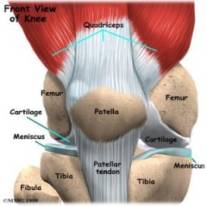
The knee is one of the joints that bear the most body weight (3). The knee is constructed of bones (1), ligaments, and cartilage (2) as shown in figure 1. These bones include the femur (thigh bone), the tibia (larger lower leg bone), fibula (other lower leg bone), and patella (knee cap).
Figure 1: Anatomy of the right knee
Ligaments function to stabilize the various motions of the knee. The two main ligaments in the knee are the ACL, Anterior Cruciate Ligament, and the PCL, Posterior Cruciate Ligament, which control the front to back motion of the knee joint. The Medial Collateral (MCL) and the Lateral Collateral Ligaments (LCL) prohibits extreme lateral movement of the joint. Ligament placement in reference to the rest of the knee joint is shown in Figure 2.
Figure 2: Anatomy of knee in flexion and extension
As illustrated in the above and below figures the cartilage is located between the femur and tibia and functions as a lubricator and shock absorber within the joint. The main cartilages of the knee, the articular and the meniscus cartilage, have important functions relative to the whole. The articular cartilage functions to provide cushion and to reduce the friction between connecting bones. The meniscus cartilage has three major functions: stability, lubrication and nutrition, and shock absorption (4).
Osteoarthritis causes knee cartilage, which normally acts as a shock absorber, to become stiff and degenerate as the joint moves making the knee more susceptible to damage. Over time, the cartilage eventually wears away causing severe pain and trauma due to the grinding of the Femur and Tibia at the point of contact. Statistics show that this disease affects over 16 million men and women as the chances of development increase with age.
Osteoarthritis is one of the most common types of arthritis. This disease is associated with the breakdown of cartilage and can be found in the hips, spine, fingers, toes, as well as the knee. Cartilage is a firm, flexible, rubbery material that can be found at the intersections of bones and acts as a natural “shock absorber.” Cartilage is made of mostly water, and has a unique ability of changing shape when it is compressed.
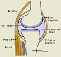
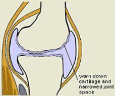
Figure 3: Normal knee cartilage (to the left) and Cartilage affected by Osteoarthritis
Osteoarthritis causes the
cartilage of joints to become stiff and inelastic, making it susceptible to
damage, deterioration, and the loss of its shock absorbing ability as shown in
figure 3. As the cartilage wears
away, it causes the tendons and ligaments in the knee to stretch causing severe
pain. If Osteoarthritis isn’t
treated, it could eventually lead to full deterioration of the cartilage causing
the femur and the tibia to rub together.
Ultimately, the best way to alleviate the symptoms of this condition is
to undergo surgery to replace the worn-away cartilage, but for people who choose
not to undergo surgery, there are other alternatives, such as living in
continuous pain, taking medications, or the most common alternative of knee
bracing.
.gif)